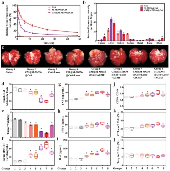
Figure 3.

Anti‐tumor effects and immune responses after combined PDT and magnetic hyperthermia with CM@M‐MON@Ce6. a) The blood circulation time, and b) biodistribution of CM@M‐MON@Ce6 in MCF‐7 tumor‐bearing mice. c) Representative images of lung tissues with observable metastatic nodules. d) The number of pulmonary metastatic nodules and e) primary tumor weights of 4T1 tumor‐bearing mice from each group over 21 d. After 5 d of combined PDT and magnetic hyperthermia, serum and primary tumor tissue were collected for the analysis of f) HMGB1, g) TNF‐α, h) IFN‐γ, and i) IL‐6 levels in serum and for the analysis of j) the ratios of CD8+T cells/CD4+T cells, k) CTL content, and l) Treg content in the primary tumor tissues. The data are presented as the mean ± S.D. (n = 5, *p < 0.05 compared with the CM@M‐MON@Ce6+Laser+ACMF group).
