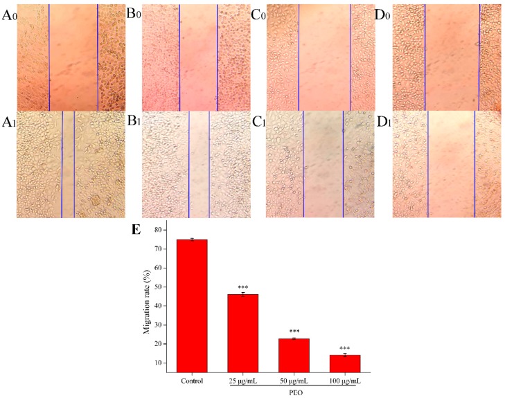
Figure 3.
Effects on MGC-803 cell migration capacity. The A–D represented PEO concentrations were 0, 25, 50, and 100 µg/mL, the subscript 0 and 1 that represented the photos (100 ×) were taken after MGC 803 cells exposing to the PEO 0 and 24 h, respectively; E was the migration rates of experimental groups that treated 24 h with PEO compared with 0 h (*** p < 0.001).

