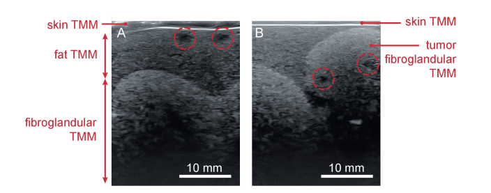
Fig. 9.
B-mode US images of the phantom acquired at two different positions. A) shows the phantom’s layered architecture and the undulating fat-fibroglandular boundary. B) shows the tumor model (made from fibroglandular TMM) embedded in the fat TMM layer, on top of fibroglandular TMM. Blood vessels are encircled.

