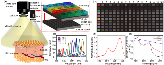
Fig. 1.
(a) Schematic of the hyperspectral imaging system that consists of a light source and a 16-channel hyperspectral camera, with wavelength bands at each channel shown in the top left. (b) The manufacturer’s data of wavelength-dependent sensitivity for 16 channels in the hyperspectral camera. (c) Waveband-based multispectral images (right 16 columns, representing 16 sub-spectral channels) of self-printed color chart as shown in the left most column, captured by the hyperspectral camera. (d) Spectral power distribution of white light source that is used in this study. (e) Absorption spectra of oxyhemoglobin (oxyHb), deoxyhemoglobin (deoxyHb) and melanin.

