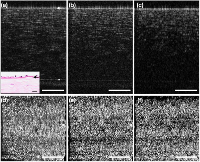
Fig. 5.
The impact of OCT axial resolution on en face visualization of corneal endothelial cell map. (a) Representative OCT B-scan of the Monkey cornea with the original axial resolution (∼ 2 µm in tissue). (b) The same B-scan with the axial resolution reduced by half (∼4 µm in tissue) and (c) quarter (∼ 8 µm in tissue). (d)–(f) The en face corneal endothelial cell images generated from the MP of the endothelial layer stacks of the respective volumes with different axial resolutions. Inset: histology slide of the cornea. The star (*) indicates the Bowman’s layer, and the arrow indicates the Descemet’s membrane. Scale bar: 200 µm

