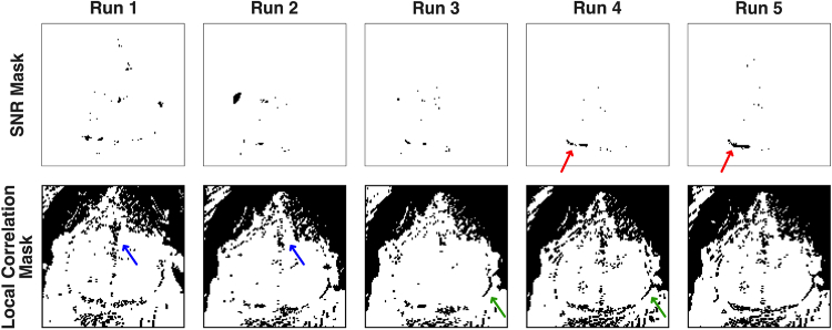
Fig. 5.
Variation in the masks created by the pixel-wise quality metric across multiple runs in the same mouse (mouse 1, sessions 1-5, prior to affine transformation). In this mouse, camera saturation was never present; so, that mask is not shown. Note also, that the mouse was repositioned after Run 1; so that run is slightly shifted relative to the others. For the SNR mask (upper row), an area at the posterior edge of the left visual cortex (red arrow) is masked in most runs, possibly due to pooling mineral oil (used in this mouse) at that location. Additionally, scattered pixels in the center of the field-of-view are excluded in each run. For the local correlation mask (lower row), the masks area are very similar across runs. Common areas excluded were the areas of fur in the upper right and upper left corners, the venous sagittal sinus (blue arrows), and along the brain-skin interface (green arrows).

