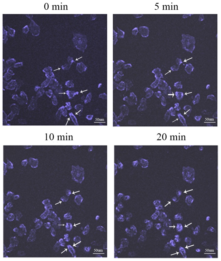
Figure 3.

Movement of IGF-1R. The distribution status of IGF-1R on the cell membrane was recorded by live staining technology. The movement track of IGF-1R can be observed with the laser scanning confocal microscopy at 0, 5, 10 and 20 min. IGF-1R, insulin-like growth factor-1 receptor. Scale bar, 50 µm.
