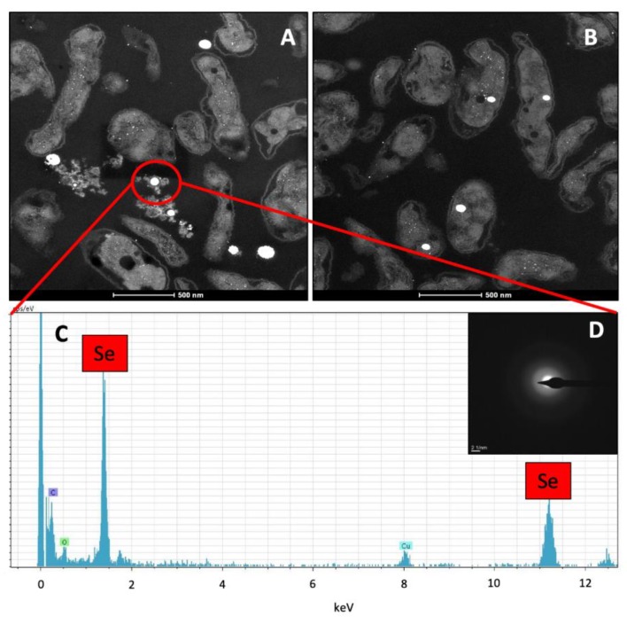
Figure 5.
HAADF-STEM analysis of an ultrathin-sectioned S. bentonitica sample incubated anaerobically at a neutral pH in the presence of 2 mM SeIV (A,B). EDX analysis revealed the Se composition of the electron-dense nanospheres (C) located both intracellular and extracellularly (A,B). SAED pattern derived from an individual Se granule (D). Scale bars: 500 nm (A,B).

