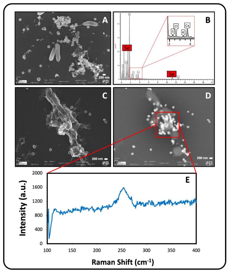
Figure 6.
VP-FESEM micrographs of Se nanospheres located extracellularly, intracellularly and attached on lysed cells of S. bentonitica (A,C,D) produced anaerobically at an initial pH of 10. EDX analysis showing the Se and S composition of the nanospheres (B). Raman analysis derived from Se nanosphere accumulations (E). Scale bars: 200 nm (A,C,D).

