
Figure 1.
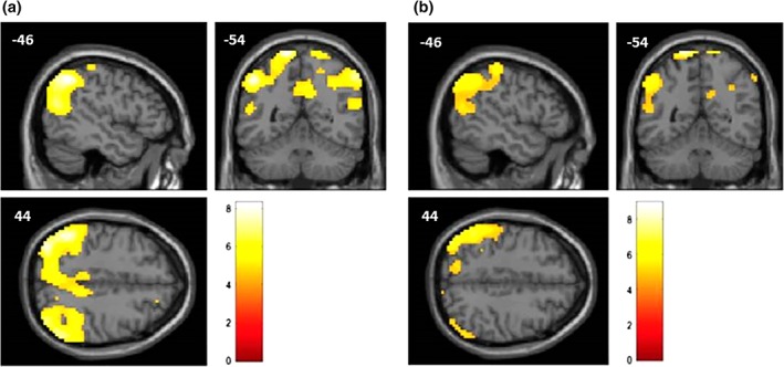
Representation, on a standard structural magnetic resonance (MR) image, of the cortical regions showing a significant metabolic decrease (measured with FDG‐PET) in the typical group (a) and the TPJ subtype group (b) compared to the control group, using the age as a nuisance variable. The regions, represented in the MNI space, are mostly posterior associative cortices. Color scale represents t‐value ((a, degree of freedom = 36; b, degree of freedom = 42)