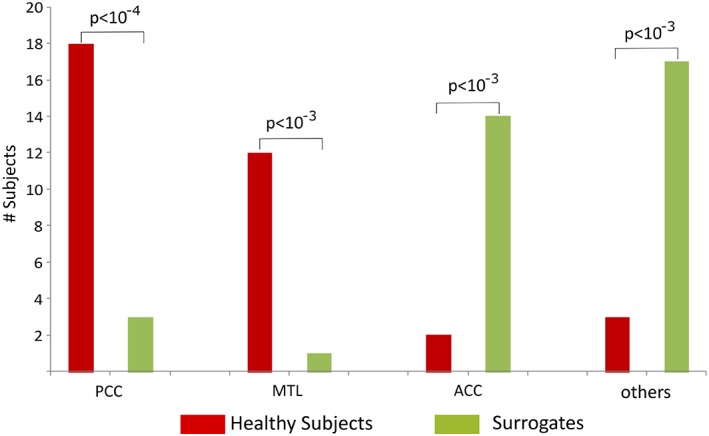
Figure 2.

Comparison of the number of subjects and surrogate data whose strongest outflow was the PCC, medial temporal lobe regions (MTL, i.e., either amygdala or hippocampus or parahippocampus), ACC, or any of the remaining 72 regions (“other ROIs”) [Color figure can be viewed at http://wileyonlinelibrary.com]
