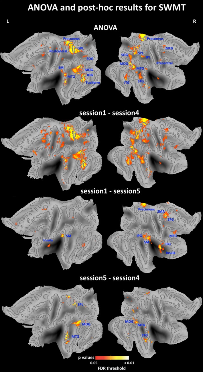
Figure 5.

ANOVA and posthoc imaging results for the SWMT. F contrast and posthoc t contrast were specified in the SwE contrast manager. The threshold was set at pFDR <0.05 corrected for multiple comparisons. Statistical maps were projected onto the flattened left and right hemisphere of the population average landmark‐ and surface‐based atlas in CARET software. Abbreviations: IFG, inferior frontal gyrus; IOG, inferior occipital gyrus; IPL, inferior parietal lobule; ITG, inferior temporal gyrus; L, left; MFG, medial frontal gyrus; MOG, middle occipital gyrus; MTG, middle temporal gyrus; R, right; SFG, Superior frontal gyrus; SM, supramarginal gyrus; SMA, supplementary motor area; SOG, superior occipital gyrus; SPL, superior parietal lobule; STG, superior temporal gyrus; SWMT, Sternberg working‐memory task [Color figure can be viewed at http://wileyonlinelibrary.com]
