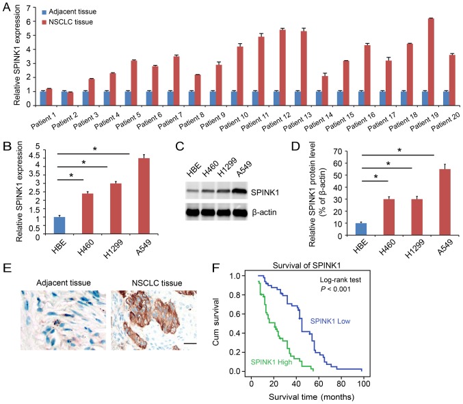
Figure 1.
SPINK1 is expressed at a higher level in NSCLC. (A) SPINK1 expression is higher in tumor samples compared with adjacent normal tumor samples. (B) The mRNA expression of SPINK1 in cell lines. SPINK1 was upregulated in NSCLC cell lines compared with HBE. (C) The protein level of SPINK1 in NSCLC cell lines. SPINK1 was upregulated in NSCLC cell lines compared with HBE. (D) Quantification of SPINK1 expression. (E) Representative images of IHC staining of SPINK1 in NSCLC and adjacent tissues. IHC showed a higher level of SPINK1 expression in NSCLC tissue samples (brown color). (F) Kaplan-Meier analysis indicated a higher level of SINK1 expression predicted a poorer overall survival time. *P<0.05 vs. control. IHC, immunohistochemistry; SPINK1, serine protease inhibitor Kazal-type 1; NSCLC, non-small cell lung cancer.

