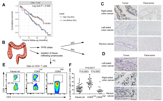
Figure 2.
CDK9 is negatively associated with the infiltration of CD8+ T cells in colorectal cancer. (A) Kaplan-Meier curves for CD8+ T cell infiltration in colorectal cancer. Levels were divided into low and high levels (cut-off, 50%). The P-value was calculated using a log-rank test. (B) Experimental scheme. (C) Immunohistochemistry of CDK9 expression in stage III colorectal cancer tissues and adjacent non-cancerous tissues. Scale bar, 50 µm. (D) Immunohistochemistry of CDK9 expression in stage IV colorectal cancer tissues and adjacent non-cancerous tissues. Scale bar, 50 nm. (E) Flow cytometric analysis of tumor-infiltrating CD8+ T cells in stage III–IV colorectal cancer tissues and paired normal tissues. (F) Frequency of tumor-infiltrating CD8+ T cells in stage III–IV colorectal cancer tissues and paired normal tissues. Each dot represents data generated from one patient (n=35). CDK9, cyclin-dependent kinase 9; IHC, immunohistochemistry; FFPE, formalin-fixed and paraffin-embedded; FC, flow cytometry; Paired ctrl, paired normal control tissues; CDK9Low T, tumor tissues with low expression of cyclin-dependent kinase 9; CDK9High T, tumor tissues with high expression of cyclin-dependent kinase 9.

