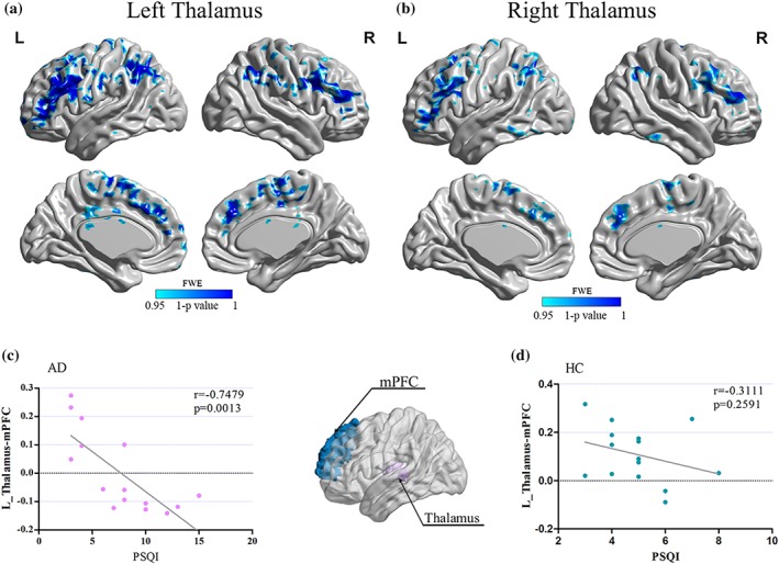
Figure 3.

(a, b) The resting‐state functional connectivity (RSFC) of thalamic differences between alcohol‐dependent patients and controls. (c, d) The correlation between functional connectivity (medial prefrontal cortex–thalamus) and PSQI score. Significantly lower resting‐state functional connectivity (RSFC) showed between left thalamus and several regions in alcohol‐dependent patients (a, FWE corrected, p < .05), that is, bilateral medial prefrontal cortex (mPFC), orbitofrontal cortex (OFC), anterior cingulate cortex (ACC), left angular gyrus, and right caudate. In addition, the right thalamus exhibited decreased RSFC with bilateral mPFC, OFC, and left caudate in alcohol‐dependent patients (b, FWE corrected, p < .05). Significant negative correlation (r = .7479; p = .0013) was found between the left medial prefrontal cortex–thalamus RSFC strength and PSQI score in alcohol‐dependent patients (c) [Color figure can be viewed at http://wileyonlinelibrary.com]
