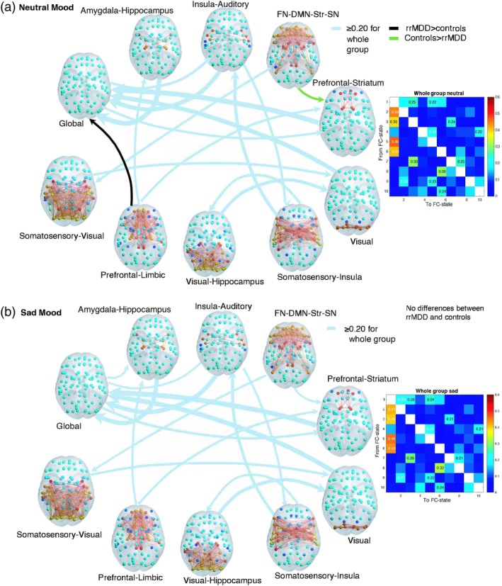
Figure 6.

(a,b) Switching probabilities for the whole group and differences between rrMDD and controls, for (a) neutral and (b) sad mood. Switching probabilities for the whole group are shown above a threshold of 20% probability of switching to show more frequent switches. The whole group switching matrices (titled “whole group neutral” and “whole group sad”) indicate the probability of, being in a given functional connectivity (FC) state (rows), transitioning to any of the other states (columns) for the whole group. FC states are represented in the cortical space, where functionally connected brain areas (represented as spheres) are colored alike. The spheres colored in yellow/red represent areas that are all positively correlated between them, but negatively correlated with the rest of the brain (cyan/blue colored spheres). The light blue arrows from and to the FC states indicate the whole group switching probabilities, scaled to the magnitude of probability of switching. Significantly different transitions after correcting for k = 10 (p < 0.05/10) for rrMDD compared to controls are illustrated in this figure, with the black arrow representing the transition that occurs with higher probability in rrMDD and in green the one that occurs with higher probability in controls (note these arrows have not been scaled according to magnitude of probability of switching). Transitioning differences between groups were calculated using a permutation‐based two sample t test with 10,000 permutations. rrMDD patients showed a lower probability of switching from the FN‐DMN‐Str‐SN state to the prefrontal–striatum state (16.5 vs. 23%, p = 0.004), and a higher probability to switch from the prefrontal–limbic state to the global state (6 vs. 1%, p = 0.003). We identified no significant between group differences in sad mood after correction for k = 10 (p > 0.05/k). Abbreviations: DMN = default mode network; FN = frontal network; MDD = major depressive disorder; rrMDD = remitted recurrent MDD; SN = salience network; Str = striatum [Color figure can be viewed at http://wileyonlinelibrary.com]
