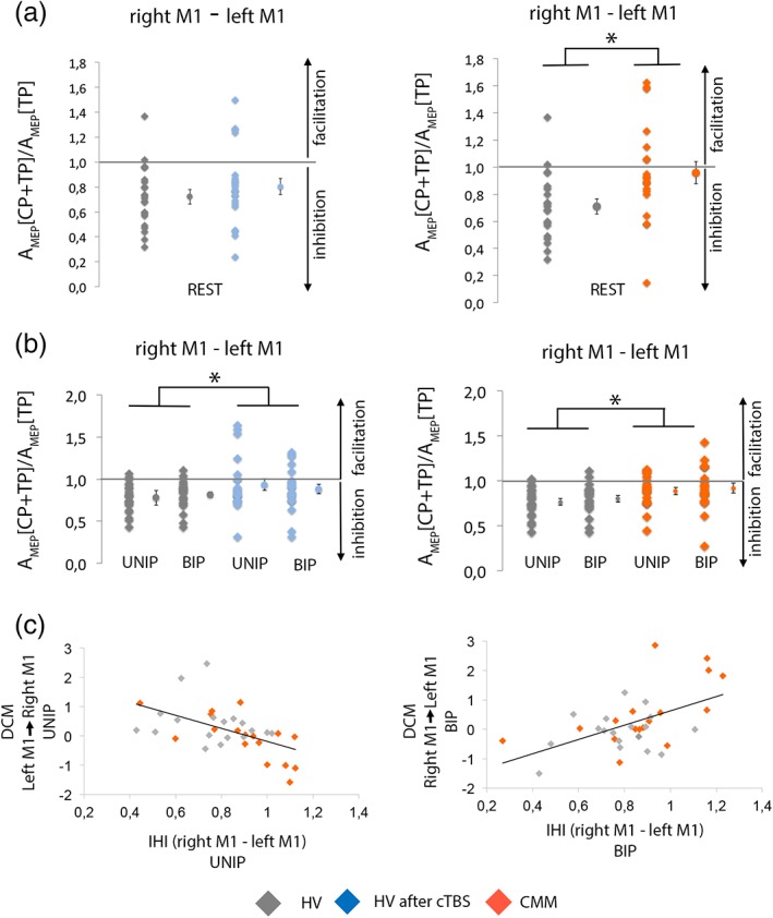
Figure 5.

Measure of the interhemispheric interactions at rest and during movement preparation and correlations between fMRI and transcranial magnetic stimulation (TMS) parameters. (a) Interhemispheric inhibition (IHI) at rest in healthy volunteers (HVs) before and after disruption of the supplementary motor area (SMA) proper (left) and in congenital mirror movement (CMM) patients and HVs (right). (b) IHI during preparation of right unimanual or bimanual movement in HVs before and after disruption of the SMA proper (left) and in CMM patients and HVs (right). Results showed that continuous theta‐burst stimulation (cTBS) in the HVs diminished the IHI from right M1 to left M1 during unimanual and bimanual movement preparation but not at rest. The IHI from the right M1 to the left M1 is decreased in CMM patients as compared to HVs both at rest and during movement preparation. Individual data are presented as dot plots alongside the mean and SEM. (c) In HVs and CMM patients, lower connectivity between left M1 and right M1 during unimanual preparation (UNIP) correlates with the degree of IHI during UNIP (left). Greater connectivity between right M1 and left M1 during BIP correlates with the degree of IHI during BIP (right) [Color figure can be viewed at http://wileyonlinelibrary.com]
