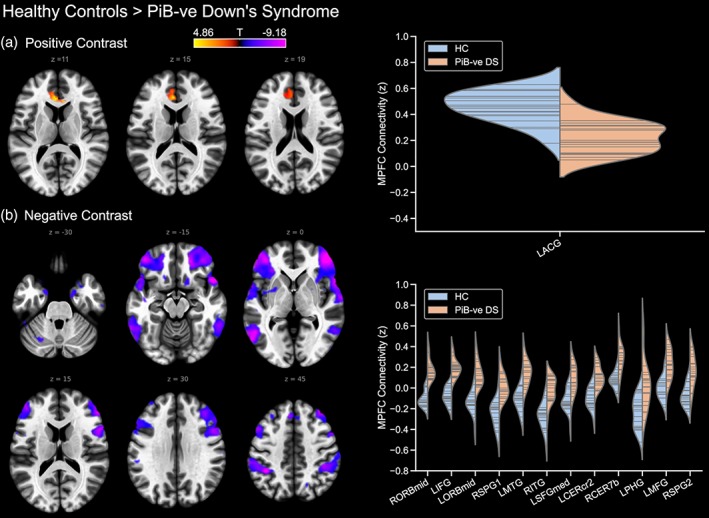
Figure 3.

Differences in default mode network connectivity between typically developing controls and PiB‐negative participants with Down's syndrome, showing (a) controls > PiB −ve Down's syndrome, and (b) PiB −ve Down's syndrome > controls. PiB −ve, PiB‐negative; where prefixed to the abbreviation of a brain region, L and R indicate the left and right hemisphere, respectively. Furthermore, a suffix of 1 or 2 to an abbreviation serves to differentiate different significant clusters located within a single brain region. ACG, anterior cingulate gyrus; CERcr2, cerebellum crus 2; CER7b, region 7b of the cerebellum; ITG, inferior temporal gyrus; MFG, middle frontal gyrus; MTG, middle temporal gyrus; ORBmid, middle orbitofrontal gyrus; PHG, parahippocampal gyrus; SPG, superior parietal gyrus; SFGmed, medial superior frontal gyrus [Color figure can be viewed at http://wileyonlinelibrary.com]
