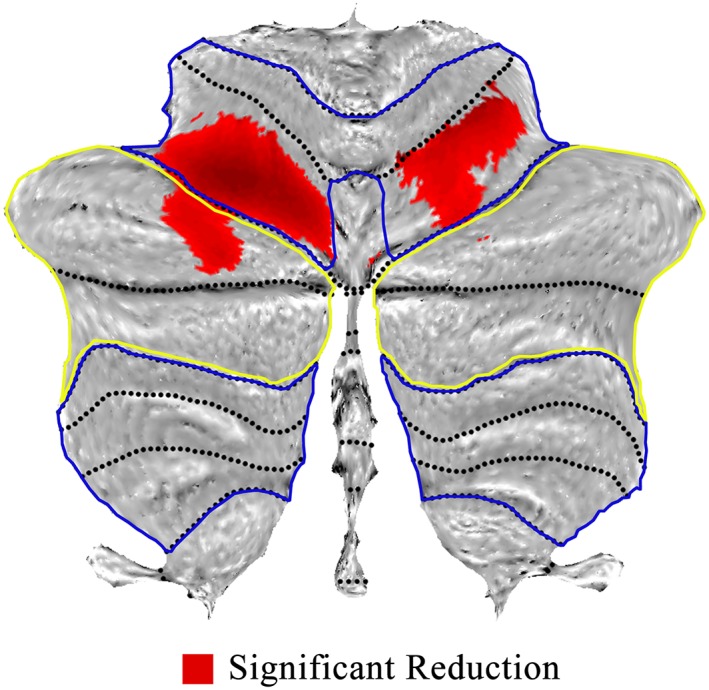
Figure 1.

Voxel‐based morphometry showing difference of gray matter within cerebellum for schizophrenia. Areas of significant reduction of gray matter (red) in the motor (within blue circle) and cognitive (within yellow circle) cerebellar territories, for patients versus healthy controls [Color figure can be viewed at http://wileyonlinelibrary.com]
