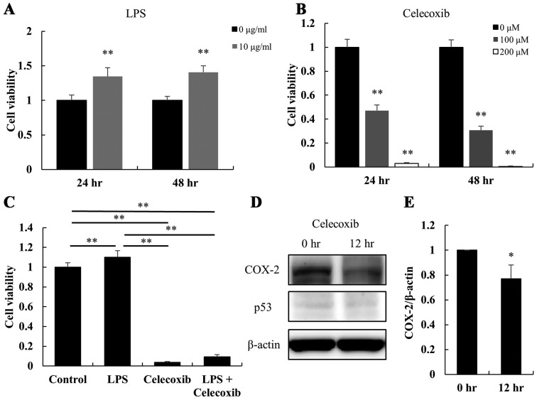
Figure 1.
Effect of LPS and celecoxib on the viability of HSC-3 cells. The viability of HSC-3 cells was determined using an MTS assay. (A) Cell viability 24 and 48 h after P. gingivalis' LPS treatment (10 µg/ml). (B) Cell viability at 24 and 48 h after treatment with celecoxib (100 and 200 µM). (C) Cell viability 48 h after treatment with celecoxib (100 µM), LPS (10 µg/ml) or the combination of these two agents. (D) The expression levels of COX-2 and p53 with/without celecoxib treatment were determined by western blotting after 0 or 12 h of treatment with 100 µM celecoxib. (E) The COX-2/β-actin ratio was calculated based on the intensity of the bands in the HSC-3 cell lines. Columns represent the mean ± standard deviation. Each experiment was performed at least in triplicate. *P<0.05 and **P<0.01 vs. untreated cells. LPS, lipopolysaccharide; COX-2, cyclooxygenase-2.

