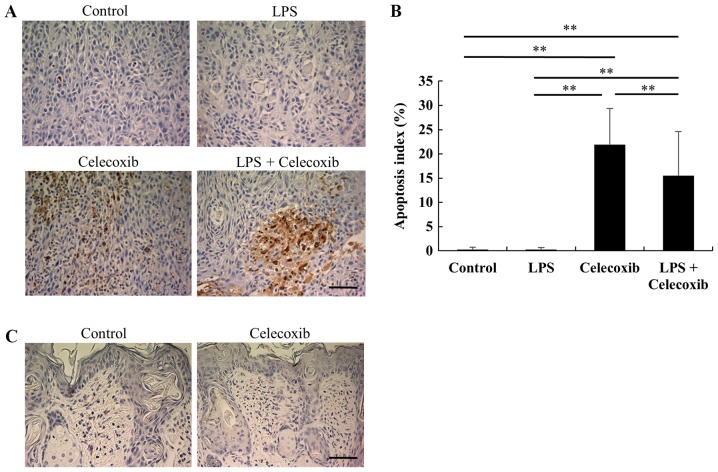
Figure 4.
Apoptosis assay in tumor xenografts in response to celecoxib with/without LPS-treatment. (A) Representative microphotographs of TUNEL staining in each group. Scale bar, 50 µm. (B) Apoptosis index by TUNEL staining. Each bar represents the mean ratio of the number of TUNEL-positive cells to the total number of tumor cells ± standard deviation. The apoptosis indices in the celecoxib-treated and LPS + celecoxib-treated group were significantly higher than those in the control and LPS-treated group, whereas the apoptosis indices in the LPS + celecoxib-treated group were significantly lower compared with those in the celecoxib-treated group. (C) Representative microphotographs of non-tumor tissues in TUNEL staining. Apoptosis was not induced in non-tumor tissues exposed to celecoxib treatment. Scale bar, 50 µm. **P<0.01 vs. control or LPS-treated group. LPS, lipopolysaccharide; TUNEL, terminal deoxynucleotidyl-transferase-mediated dUTP nick end labeling.

