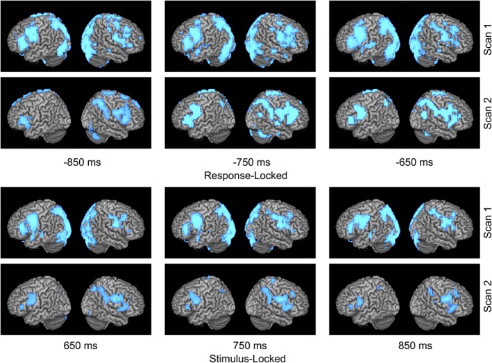
Figure 1.

Left to right shift in language laterality. Time course of language network activation over two scans for patient with a left frontal glioma who experienced a rightward shift in LI of −0.416 (0.116 to −0.3) over 1 year. Activity is time‐locked to either stimulus or response onset. Combined LI score is calculated with both stimulation and response activations. At second scan for both response and stimulus locked conditions, larger area of activation (in blue) can be observed on right hemisphere compared to less area of activation on left hemisphere [Color figure can be viewed at http://wileyonlinelibrary.com]
