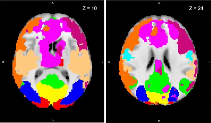
Figure 1.

Resting‐state functional magnetic resonance imaging network maps identified by Smith and Nichols (2009) overlaid onto a standard brain in Montreal Neurological Institute (MNI) space. Axial slices displayed at coordinate shown. Yellow, medial visual; red, visual occipital pole; blue, lateral visual; green, default mode; not shown, cerebellum; light blue, sensorimotor; light orange, auditory; purple, executive control; orange, right frontoparietal; dark pink, left frontoparietal [Color figure can be viewed at http://wileyonlinelibrary.com]
