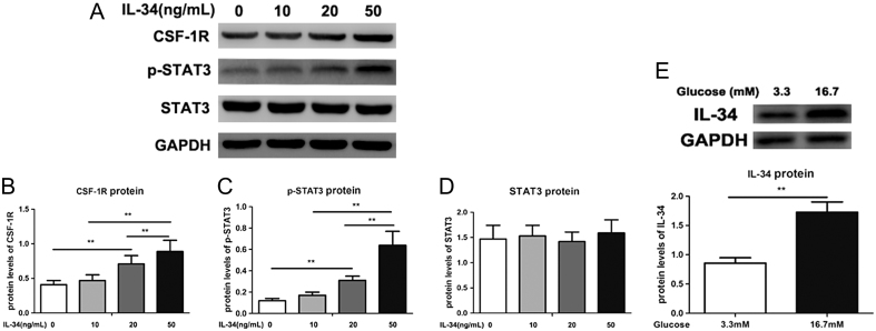
Figure 3.
Protein expression of IL-34 in pancreatic β cells cultured in the treatment of high glucose and CSF-1R contributes to the biological activities of IL-34. (A) The protein expression levels of CSF-1R and pSTAT3/STAT3 were measured by Western blotting. The ratios of CSF-1R/GAPDH (B), p-STAT3/GAPDH (C) and STAT3/GAPDH (D) were determined to give a mean net density. Protein expression of IL-34 in MIN6 cells cultured as exposure to elevating doses of glucose. (E) The protein expression levels of IL-34 were measured by Western blotting and a representative result is presented. Data are presented as the means ± s.e. **P < 0.01 was considered significant.

 This work is licensed under a