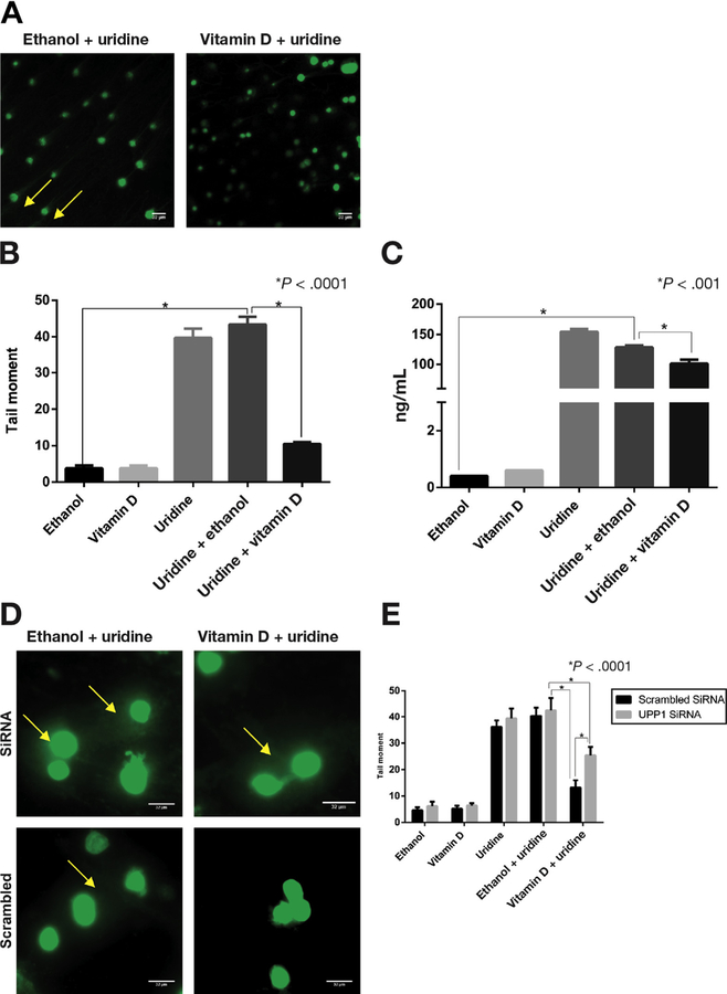
Figure 4.
Vitamin D suppression of uridine-induced DNA damage. FLARE assay was performed to examine uridine-induced dUTP incorporation in YAMC cells. (A) Electrophoresed cells stained with SYBR green after treatment with uridine plus ethanol and uridine plus vitamin D. Arrowhead points to the tail that results from DNA damage seen with uridine plus ethanol but not with uridine plus vitamin D treatment. (B) DNA damage as estimated by tail moment using ImageJ software. Representative comparisons and P values are shown on the graph for clarity. There was significantly increased tail moment with uridine alone and with uridine plus ethanol treatments (both P < .0001). There was significantly less tail moment in cells treated with the combination of uridine plus vitamin D compared with cells treated with uridine plus ethanol or with uridine alone (both P < .0001). (C) Uridine concentrations by treatment condition. Uridine concentration was significantly increased with uridine treatment (both uridine and uridine plus ethanol) (P < .0001). Uridine concentrations were lower in the cells treated with the combination of uridine plus vitamin D compared with the cells treated with uridine plus ethanol or uridine alone (P < .0001). (D) Representative FLARE results after YAMC cells were transfected with UPP1 siRNA or scrambled siRNA and treated with uridine plus ethanol and uridine plus vitamin D. Arrowheads point to the tails resulting from DNA damage. (E) Tail moments were quantified using ImageJ. Uridine plus vitamin D treatment showed significantly greater tail moment in UPP1 siRNA transfected cells compared with scrambled control (P < .0001).

