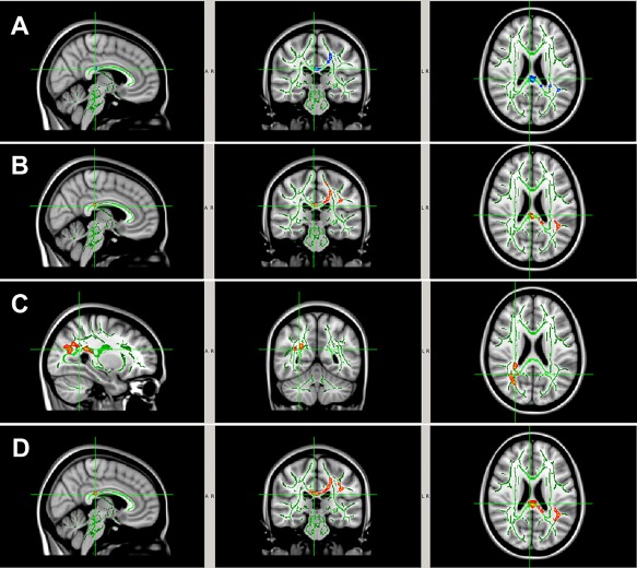
Figure 4.

WM regions with significantly larger pre‐ to post‐season DTI change ((A) larger FA increase; (B) larger MD decrease; (C) larger AD decrease; (D) larger RD decrease) in the noncollar group (n = 10) than that in the collar group (n = 13) in Season 1. The areas with significant group difference of longitudinal DTI change include body and splenium of corpus callosum, internal capsule, superior and posterior corona radiata, posterior thalamic radiation (including optic radiation), and superior longitudinal fasciculus. [Color figure can be viewed at http://wileyonlinelibrary.com]
