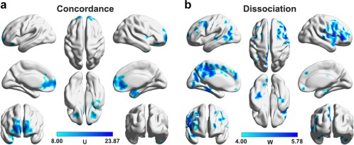
Figure 11.

Regions that simultaneously exhibited decreased DC and CBF (i.e., concordance, a) and that only exhibited decreased CBF (i.e., dissociation, b) in MDD. Multiple regions were identified to only show decreased CBF but have intact DC in the patients that mainly included the dorsal prefrontal, posterior medial parietal, and occipital cortices [Color figure can be viewed at http://wileyonlinelibrary.com]
