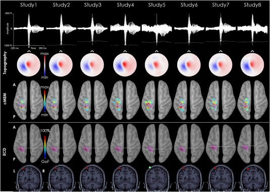
Figure 6.

Patients with focal cortical dysplasia in the left postcentral gyrus. The MEG signal of the average IEDs time‐course is pretty consistent across studies. MEG topography at the peak is also very stable across studies. Whereas dMSI always recovers the interictal source with an estimation of its extent in the left postcentral gyrus, ECD localization corresponds to the same area, but the dipole is very close to the skull or even outside it. The pink cortex corresponds to the epileptic focus as assessed by iEEG. [Color figure can be viewed at http://wileyonlinelibrary.com]
