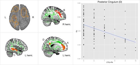
Figure 2.

Displayed are fractional anisotropy images of all subjects (n = 114), which are projected onto a skeletonised image representing the centres of all tracts common to the group. Images are fully corrected for multiple comparisons using threshold‐free cluster enhancement at p < .05. The color‐coded areas (subfigure 1–4) depict the negative correlation between the physical neglect subscale of the CTQ in the bilateral anterior thalamic radiation around the middle frontal gyrus, which included the forceps minor and unciate fasciculus (area A) on the left side and the inferior fronto‐occipital fasciculus on the right side (area B). The regions also include the left inferior fronto‐occipital fasciculus and inferior longitudinal fasciculus (area C), and the posterior cingulum near the precuneus (area D). All regions were voxelwise correlated to CTQ‐PN, but only FA in the posterior cingulum mediated trait anxiety (subfigure 5, also see Figure 3) [Color figure can be viewed at http://wileyonlinelibrary.com]
