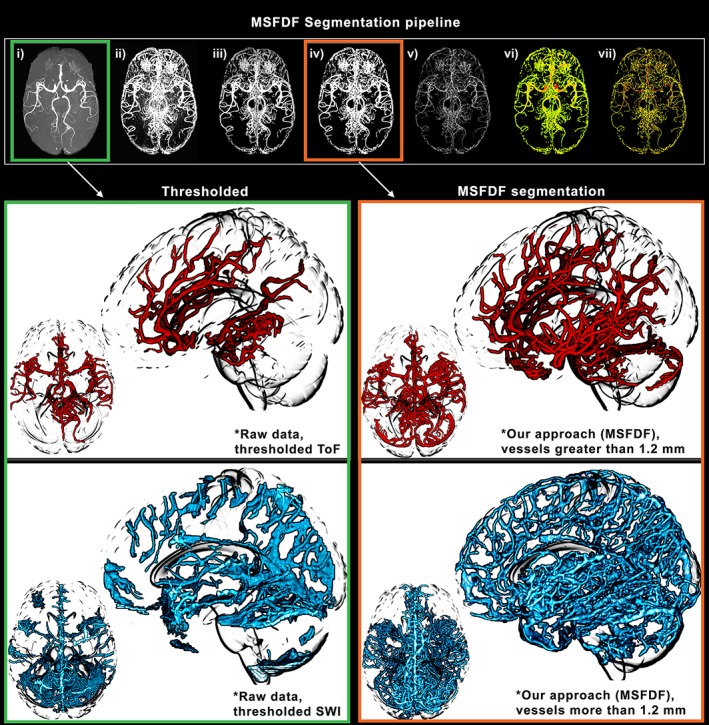
Figure 1.

Illustration of our vessels segmentation pipeline. The extraction of small vessels (~1 mm) necessitates a complex pipeline of image processing due to inconsistencies in signal‐to‐noise ratio and intensity leveling across regions of the brain. (a–g) Each step of the segmentation and diameter extraction explained in the Methods section is shown here. Green: manual thresholding of a ToF and SWI image from a single subject. Orange: vessels extracted using our multiscale Frangi diffusion filter (MSFDF) pipeline on the same ToF
