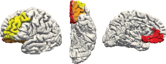
Figure 1.

Cortical regions extracted from Freesurfer ROIs for a sample participant's left hemisphere, showing lateral (left), inferior (middle), and medial (right) views. Regions selected are the dorsolateral prefrontal cortex (yellow), medial prefrontal cortex (red), and orbitofrontal cortex (orange) [Color figure can be viewed at http://wileyonlinelibrary.com]
