
Figure 2.
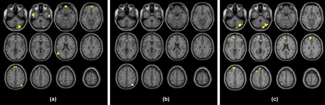
Clusters resulting from the FD > HC between‐group contrast for the seed placed at level of the MPFC (a), of the ventral portion of the precuneus (b), and of the right parietal lobule (c). Results are superimposed for anatomic reference to a single individual's T1‐weighted volume in the standard MNI space, with subject's right at the observer's right. Significance for all clusters is p < .008, family wise error corrected at cluster level [Color figure can be viewed at http://wileyonlinelibrary.com]