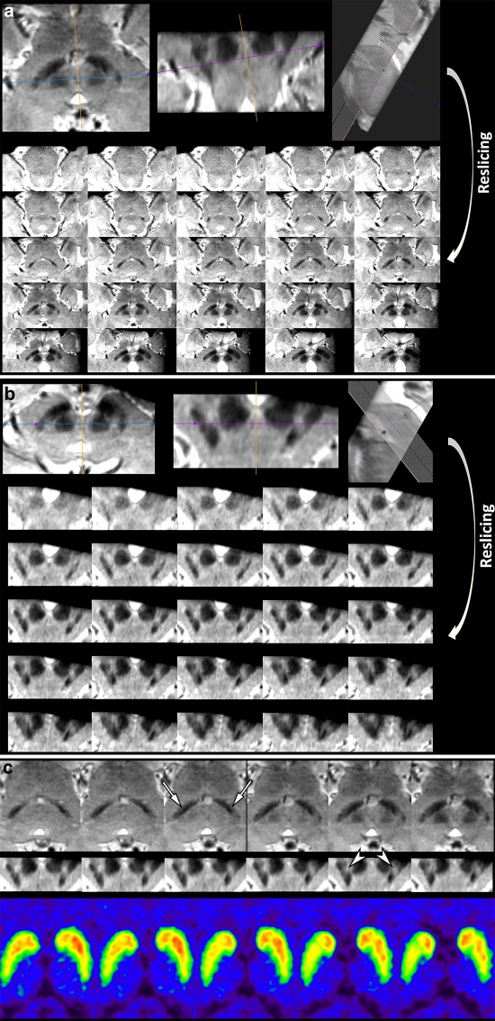
Figure 2.

Reslicing susceptibility map‐weighted images when they are not obtained symmetrically, which are caused by various technical factors such as the patient's brain shape or inadvertent positioning. (A) SMWI of a 78‐year‐old female with early‐stage IPD obtained parallel to the line from the posterior commissure: upper border of the pons was repositioned to be as symmetric as possible at the level of the lower border of the red nucleus, and thereafter the images were resliced at an increment of 0.5 mm. (B) The resliced images were reformatted perpendicular to the midbrain axis at an increment of 0.2 mm. (C) Although this was the most mal‐aligned imaging in this study, it can determine abnormality in the bilateral nigrosome 1 regions (arrows; right more affected than left) below the red nucleus, when compared to their normal features in Figure 1A. Note the putative left nigrosome 4 region is normal, while right nigrosome 4 region is possibly abnormal (arrowheads). FP‐CIT PET shows mild abnormality in the bilateral basal ganglia, right greater than left, which is well correlated with the findings on SMWI. [Color figure can be viewed at http://wileyonlinelibrary.com]
