
Figure 6.
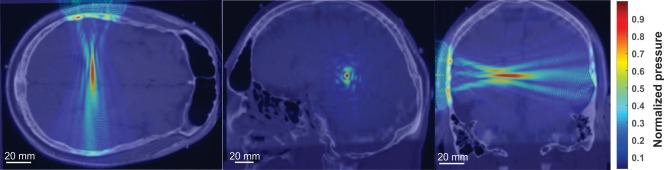
Acoustic modeling of ultrasound wave propagation. Normalized pseudocolor pressure distribution in a single subject acoustic model of ultrasound wave propagation using CT scan to account for the effect of skull morphology. Image shows MRI with CT overlay. Transducer is placed at the top of the transverse image (left) and on the left of the coronal image