
Figure 1.
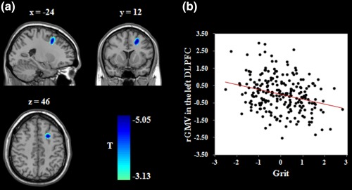
Brain regions related to grit. (a) Brain images depicting the negative association between grit and the rGMV in the left DLPFC. (b) Scatter plot showing the correlation between grit and left DLPFC volume (r = −.27, p < .001). Age, gender, total GMV, and general intelligence were adjusted for in these analyses. rGMV = regional gray matter volume; DLPFC = dorsolateral prefrontal cortex [Color figure can be viewed at http://wileyonlinelibrary.com]