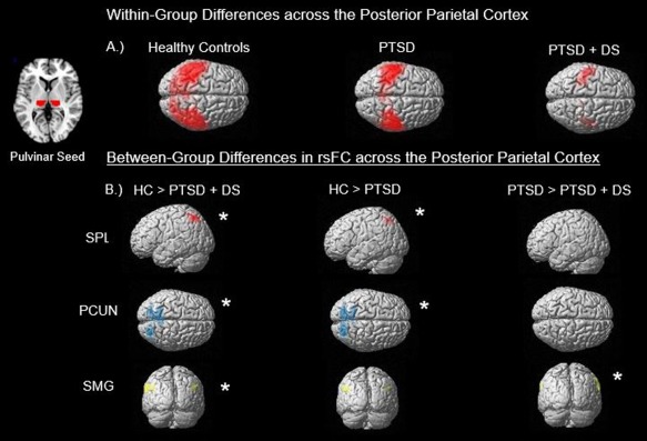
Figure 1.

This illustration summarizes the main findings from the (a) within‐group and (b) between‐group analyses. Section (a) demonstrates the resting‐state functional connectivity (rsFC) across all subjects within their respective group restricted to the posterior parietal cortex. Also included is a generated mask of the pulvinar which was used as the seed to which whole‐brain voxel correlations were conducted against. As seen, healthy controls (HC) demonstrate the greatest pulvinar rsFC and PTSD + DS had the least, with PTSD showing an intermediary of the two. Section (b) breaks down the findings of the between‐group contrasts to three regions, the superior parietal lobule (SPL), displayed in red, precuneus (PCUN), displayed in blue, and supramarginal gyrus (SMG), which is contained within the inferior parietal lobule (IPL), displayed in yellow. Findings were restricted to the left SPL, as it was the only hemisphere displaying any results. Additionally, significant results for the PCUN were generated for both the left and right hemisphere in HC > PTSD + DS, and only the left hemisphere in HC > PTSD. Last, significance for PTSD > PTSD + DS was found within the right SMG. Asterisks indicate significance at pFWE < .05, k = 10 [Color figure can be viewed at http://wileyonlinelibrary.com]
