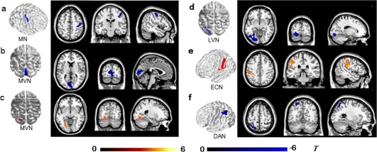
Figure 3.

Brain regions with significant differences in intra‐network functional connectivity (FC) between stroke patients and controls (a–f). Warm and cold colors indicate regions with higher or lower intranetwork FC in stroke patients compared with healthy controls (p < .05, AlphaSim corrected). The colored bars indicate the T values of the two‐sample t test. MN, motor network; MVN, medial visual network; LVN, lateral visual network; DAN, dorsal attention network; ECN, executive control network [Color figure can be viewed at http://wileyonlinelibrary.com]
