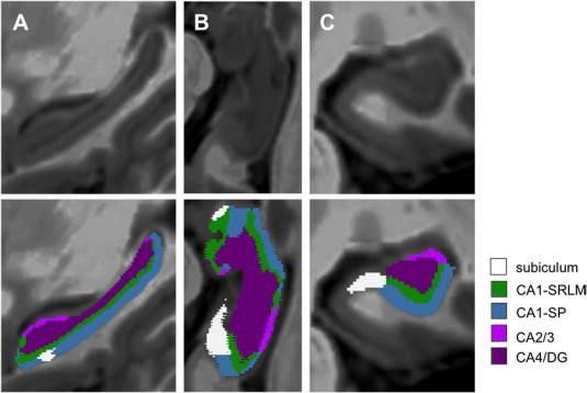
Figure 1.

Segmentation of the five hippocampal subfields. Super‐resolution T1‐weighted images (0.5 × 0.5 × 0.5 mm3) centered on the left hippocampus of a patient with clinically isolated syndrome (PwCIS) in the sagittal plane (a), in an oblique axial cut parallel to the plane of the hippocampus (b) and in the coronal plane (c). Five hippocampal subfields were automatically segmented (and manually corrected if needed) according to the atlas of Winterburn et al.: the subiculum, the stratum pyramidale of CA1 (CA1‐SP), the stratum‐lacunosum‐moleculare of CA1 (CA1‐SRLM), CA2/3, and CA/4dentate gyrus (CA4/DG) [Color figure can be viewed at http://wileyonlinelibrary.com]
