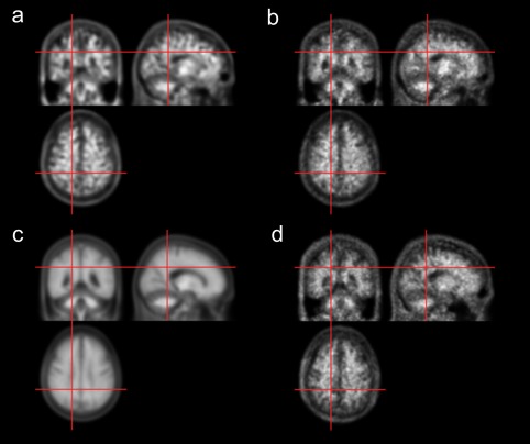
Figure 3.

11C‐PIB PET templates generated using deep neural networks for an amyloid negative case. (a) CAE‐generated template. (b) GAN‐generated template. (c) Average template. (d) Spatially normalized image using MRI, which serves as the label for CAE and GAN training [Color figure can be viewed at http://wileyonlinelibrary.com]
