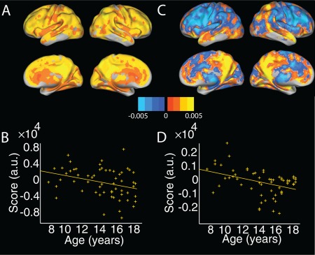
Figure 2.

Two age‐associated principal components in CBF. (a) Spatial distribution and (b) associated scores plotted against age for the top principal component, accounting for 40.6% of the variance. This component includes regions of high intensity in the posterior cingulate/precuneus. Note component scores showed only a trend‐level association with age (P = 0.06). (c) Spatial distribution and (d) associated scores plotted against age for the fourth principal component, accounting for 3% of the variance. This component distinguished medial and lateral parietal cortex from dorsal/lateral prefrontal regions. CBF = cerebral blood flow. [Color figure can be viewed at http://wileyonlinelibrary.com]
