
Figure 4.
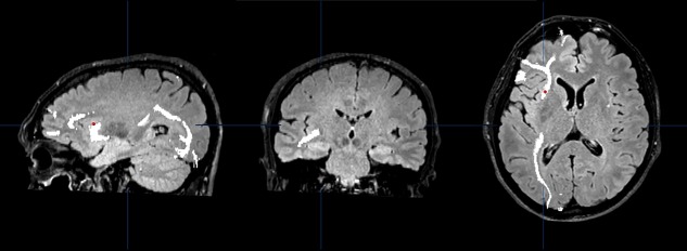
Oligodendroglioma in the left frontal lobe (patient N. 9). Sagittal, coronal and axial view of the trajectories of the left inferior fronto‐occipital fascicle (IFOF, in white) in the DTI study acquired before resection. DES applied over the IFOF did not interfere with digit span. The site of stimulation is marked with a red dot registered through the neuronavigation system. [Color figure can be viewed at http://wileyonlinelibrary.com]