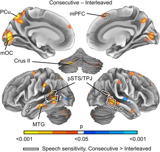
Figure 5.

Brain regions showing different ISC between Consecutive and Interleaved conditions. The data are thresholded at P < 0.05, FWER corrected. Warm colors correspond to areas showing increased ISC in Consecutive and cold colors increased ISC in Interleaved condition. White line indicates areas showing different sensitivity to speech in Consecutive vs. Interleaved condition in Figure 7. Abbreviations: mOC, medial occipital cortex; mPFC, medial prefrontal cortex; MTG, middle temporal gyrus; PCu, precuneus; pSTS, posterior superior temporal sulcus; TPJ, temporoparietal junction.
