
Figure 5.
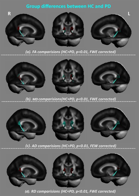
Between‐group differences of diffusion properties along the parahippocampal section. As compared with the HC group, PD patients had altered white matter mainly located in the posterior cingulum in PD patients (P < 0.01, FWE corrected) with (a) decreased fractional anisotropy (FA), (b) increased mean diffusivity (MD), and (c) increased radial diffusivity (RD). [Color figure can be viewed at http://wileyonlinelibrary.com]