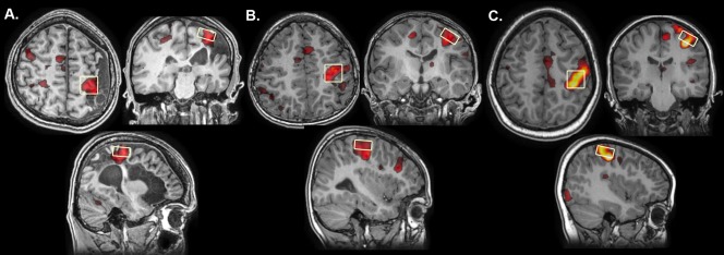
Figure 1.

Function‐guided MRS voxel placement. MRS voxel placement viewed on axial, coronal and sagittal slices approximates the hand‐knob in the precentral gyrus in a representative (A) Arterial, (B) PVI and (C) TD participant. Voxels were placed corresponding to task fMRI activations measured during a finger tapping task. Lesioned hemispheres have all been reoriented to the right side.
