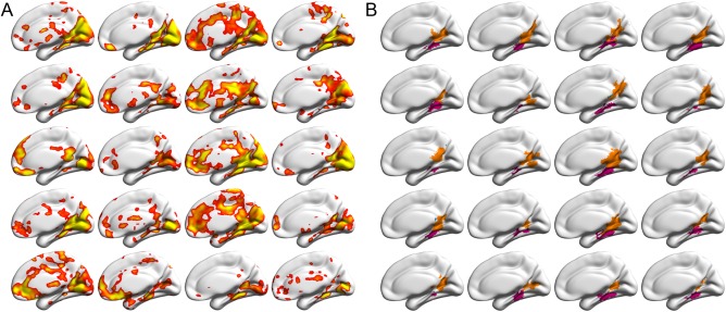
Figure 1.

Sample activations and delineated SSRs in the right hemispheres from the 20 randomly selected brains, overlaid on the standard MNI152 cortical surfaces. Because of space limitations, only the medial view is presented. For the lateral view, see Supporting Information Figure S2. (A) The individual activation maps for the contrast between scenes and objects, derived from three runs of fMRI data for each subject, uncorrected P < 0.01 (right‐tailed). (B) The delineated subject‐specific SSRs for the activations in (A). PPA and RSC were shown in magenta and orange, respectively. Each cell corresponds to one subject. Abbreviations: PPA, parahippocampal place area; RSC, retrosplenial cortex; SSR, scene‐selective region. [Color figure can be viewed at http://wileyonlinelibrary.com]
