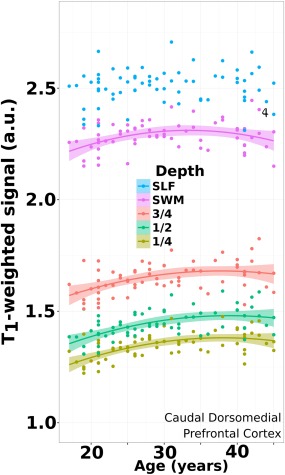
Figure 3.

Age trajectory from superficial cortex to deep WM. Data plotted for an ROI (caudal dorsomedial prefrontal cortex) in all subjects where the quadratic model fit the signal data at all depths. This is contrasted against the signal of a volumetric ROI placed in the superior longitudinal fasciculus (SLF) in deep white matter, which demonstrated no significant effect with age over this range. Shaded regions denote the 95% confidence interval. [Color figure can be viewed at http://wileyonlinelibrary.com]
