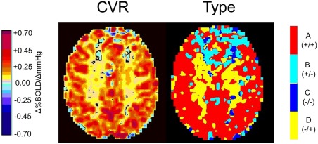
Figure 4.

Example CVR and type maps for an axial slice obtained from analysis of BOLD MRI responses to a ramp CO2 stimulus in a healthy 75‐year‐old male (c2). The colour scales are as shown. The WM in this subject shows a positive CVR whereas the type map indicates that the pattern of flow in the WM is biphasic (D −/+ type). In the frontal lobes, the flow is also biphasic (B +/− type). The biphasic aspect of both B +/− and D −/+ patterns results in small CVR values.
