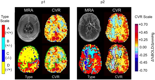
Figure 6.

Example detailed comparisons of CVR and types maps for two patients. A detailed examination of these maps is given in the discussion. For p1, the type map (bottom row left), the voxels with D −/+ are outlined (green line) and reflected onto the CVR map (green line, bottom row, right). For p2, the type map (bottom row, left) the voxels with B +/− are outlined in black, and the C −/− in purple. These are reflected onto the CVR map (bottom row, right). Note that the biphasic D −/+ are interpreted as positive and negative CVR. In p2, the biphasic B +/− are interpreted as positive and negative CVR, but the monophasic C −/− are only negative CVR, or steal.
