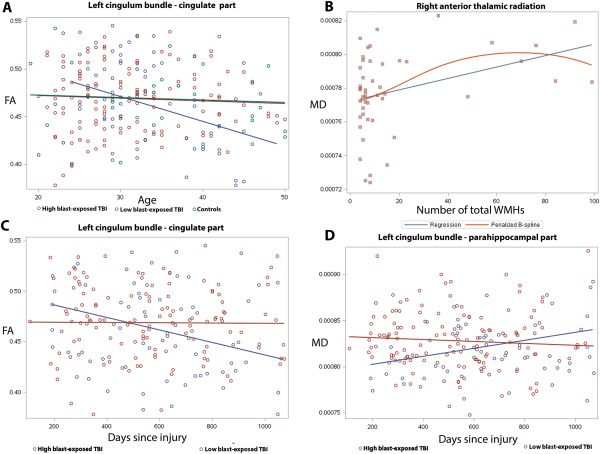
Figure 3.

Results of the interaction between blast exposure and aging on cross‐sectional FA (A), the association between MD and WMHs (B), and the interaction between blast exposure and time since most severe blast injury (C, D) on DTI measures using TRACULA ROI analyses. Color‐coding with blue, red and green corresponds to high blast‐exposed, low blast‐exposed and controls, respectively. (A) Linear models of mean FA versus age in the cingulate part of left cingulum bundle that revealed a more rapid regional cross‐sectional aging trajectory toward reduced white matter integrity (reduced FA) in the high blast‐exposed group (at least 3 blast exposures) than low blast‐exposed (1 or 2 blast exposures) and controls (P < 0.05). (B) Linear (blue) and B‐spline (red) models of mean MD of the right anterior thalamic radiation and the number of brain WMHs in the mTBI participants with large number of WMHs (larger than 3 WMHs in total) (P < 0.05). (C, D) Regional mean FA of the cingulate part of left cingulum bundle (C) and mean MD of the left parahippoocampal part of the left cingulum bundle (D) in mTBI patients shows that the trajectories toward reduced white matter tissue integrity, either decreased FA (C) or increased MD (D) with increasing time since blast injury, were unique to previous blast exposures.
