
Figure 7.
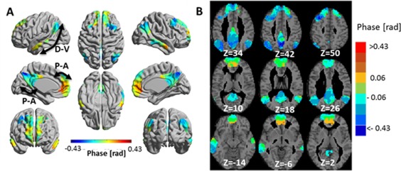
Phase 3‐seed‐SPM of the default mode network (DMN) for wavelet scale 1 (0.02–0.03 Hz). SPMs were constructed from: left medial orbitofrontal cortex, left angular gyrus and left posterior cingulate gyrus seeds. Phases were calculated with respect to the phase of the left medial orbitofrontal cortex and are shown for clusters with significant amplitude. (A) The phases in radians are projected on 3D brain templates in MNI space and indicated by the color bar. The arrows indicate the suggested temporal organizations, with D‐V: dorsal‐ventral and P‐A: posterior‐anterior. (B) Zoom into nine axial MRI slices covering most of the DMN. MNI z coordinate is indicated for each slice. [Color figure can be viewed at http://wileyonlinelibrary.com]